Laura E. Keller; Terri-Ann N. Kelly; Jacqueline M. Chevalier; Hyung Jin Jung; Garett B. Pearson; Laila Begum; Olivia S. Beane; Sarindr Bhumiratana; Lisa A. Fortier
The American Journal of Sports Medicine. 2026;54(3):622-634, published: February 10, 2026
https://doi.org/10.1177/03635465251409083
1 citation · 1 en 2025–2026
Les titres, résumés et légendes des publications sont présentés en anglais.
Abstract
Background:
Fresh osteochondral allograft (OCA) transplantation effectively repairs cartilage and subchondral bone; however, the persisting shortage of available donor OCAs and their short shelf-life make scheduling surgeries and meeting patient demand challenging. Attempts have been made to develop tissue-engineered solutions to address the limitations of OCA; nonetheless, these have failed to progress beyond the preclinical stage.
Purpose:
To assess the safety and efficacy of a tissue-engineered osteochondral allograft (TE-OCA) as compared with equine OCA in an equine osteochondral defect model.
Study Design:
Controlled laboratory study.
Methods:
Bilateral critical-size (10 × 7.0-7.5 mm) osteochondral defects were surgically created on the femoral medial trochlear ridge of healthy, skeletally mature horses (2-5 years; n = 8). A TE-OCA was placed into 1 defect, and an OCA was placed into the contralateral defect as a positive control. At surgery, throughout the study, and at sacrifice (14 months), quantitative evaluation of lameness and synovial fluid composition was obtained, while radiographs, arthroscopies (4, 10, and 12 months), or gross images, and synovial membrane were qualitatively scored. Postmortem, joints and grafts were evaluated using T1- and T2-weighted magnetic resonance imaging, quantitative computed tomography, and biomechanical testing. Histology and immunohistochemistry were performed on osteochondral blocks that were qualitatively scored.
Results:
TE-OCA exhibited better cartilage-cartilage integration on histology (97.1 ± 7.6 vs 41.7 ± 45; P = .03) and a lower T1ρ quantitative score (65.6 ± 10.6 vs 72.8 ± 5.5; P = .03) than OCA, indicative of an intact regeneration of cartilage matrix. TE-OCA matured in vivo, with biomechanical instantaneous and equilibrium compressive moduli improving to match that of OCA (1.74 ± 0.7 vs 1.96 ± 1.2 MPa [P≥.999], and 0.39 ± 0.2 vs 0.18 ± 0.2 MPa [P = .22], respectively). There were no differences between total radiographic scores (3.6 ± 2.7 vs 2.1 ± 1.7; P = .9), total International Cartilage Repair Society cartilage scores (5.1 ± 4.9 vs 2.8 ± 0.8; P = .6), and total morphologic MRI scores (8.9 ± 2.8 vs 6.2 ± 3.7; P = .3) at the study end. No off-target effects were seen.
Conclusion:
TE-OCA is comparable to OCA in multiple safety and efficacy measures of osteochondral defect repair.
Utilisation des équipements
La section de méthode mentionne un appareil d’essais Biomomentum et décrit des essais de indentation et de mesure locale, compression, friction.
Les détails du protocole sont conservés en anglais dans cette rubrique; consulter la publication originale pour le contexte expérimental complet.
Appareil : Mach-1 Mechanical Tester.
Matériaux (2)
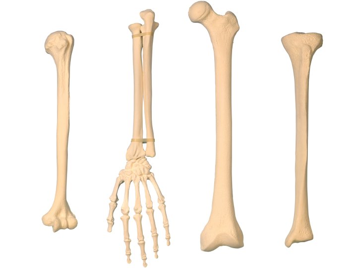
Os

Cartilage auriculaire
Configurations de test (5)

Compression Non-Confinée

Indentation (Raideur)

Friction

Cartographie 3D d'épaisseur
