Hitendra Kumar
PhD Thesis, 2021. Vancouver : University of British Columbia Library
https://doi.org/10.14288/1.0400478
Les titres, résumés et légendes des publications sont présentés en anglais.
Abstract:
The natural tissues are architecturally complex and heterogenous in nature and comprise of a dense vasculature for facilitating nutrient and chemokines transport. To mimic the functionality of natural tissues, bioprinting methods should be able to capture the tissue heterogeneity and create a vascularized scaffold. Stereolithography (SLA) bioprinting is a promising process to fabricate high-resolution anatomically accurate tissue analogues. However, non-optimized bioinks and poor design of the scaffolds limit the capability of SLA bioprinting. In view of these challenges, a method to obtain SLA bioprinting compatible gelatin methacryloyl (GelMA) bioinks which maintain a low-viscosity liquid state at room temperature is presented. The method considers GelMA synthesis parameters – solvent, pH and duration; to tune the end product. The obtained bioinks exhibit superior photopolymerization kinetics, biocompatibility and printability. Addition of a three-component photoinitiator further improved the sensitivity of the bioinks to visible light and resulted in an accelerated photopolymerization. The printing time was nearly halved and structurally stable structures could be formed with lower GelMA concentration. Next, a numerical model is developed to predict the hydrogel formation in mask photopolymerization. Considering a realistic Gaussian illumination and diffusive transport of chemical species within the bioink, the model was able to predict the printing artifacts like overcrosslinking and distortion in the hydrogel shape. In the last step, a vascular network generation method inspired from leaves and tree growth is presented. Biologically relevant vasculature was formed guided by medical image derived tissue structure and porosity. By studying nutrient transport, optimized networks with increased complexity were obtained. In combination, the described bioinks and the numerical models provide effective tools for designing the tissue mimics with embedded heterogeneity and an intrinsic vasculature to achieve the functionality of natural tissues.
Utilisation des équipements
Le Mach-1 Mechanical Tester (v500c) a appliqué une déformation sinusoïdale en cisaillement et enregistré la réponse en force d’une bioencre GelMA pendant sa photopolymérisation afin de caractériser la cinétique de photoréticulation. La sonde et la plateforme portaient des plaques de verre revêtues de TMSPMA entre lesquelles était placée la bioencre. Un projecteur DLP distinct assurait l’éclairage reproduisant le dispositif de bio-impression.
Article et documents
Consulter l’article
Matériaux (2)
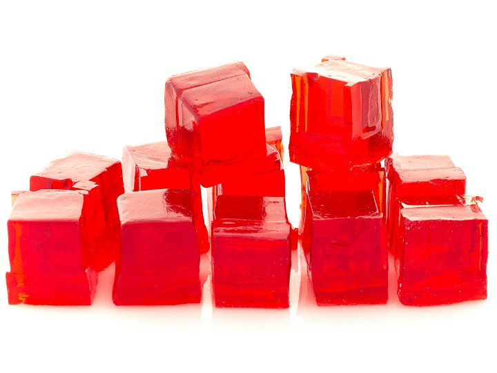
Hydrogel

Matériau par impression 3D
Configurations de test (3)

Cisaillement

Analyse Mécanique Dynamique
