Kathy K. Lee; Adele Changoor; Marc D. Grynpas; Jane Mitchell
Bone, Volume 208, 2026, 117893, ISSN 8756-3282, July 2026
https://doi.org/10.1016/j.bone.2026.117893
1 citation · 1 en 2025–2026
Les titres, résumés et légendes des publications sont présentés en anglais.
Abstract:
Gαq/11 proteins that couple G protein-coupled receptors to stimulate phospholipase C play important roles in the skeletal system as previously demonstrated by bone abnormalities when activating mutations or when overexpression of these G proteins occur in mice or humans. Here we investigated the effect of increased Gα11 in osteoblastic cells on bone fracture repair in transgenic (G11-Tg) mice in comparison to wild type (WT). Following stabilized tibial osteotomies in male mice, fracture healing was examined weekly over 4 weeks by micro-CT, histomorphometry, and gene expression analysis and bone biomechanics after 4 weeks. Histomorphometry showed diminished cartilage in G11-Tg mice at peak soft callus formation. Histology showed diminished osteoblasts on the healing bone of G11-Tg mice throughout the 4 weeks ending with lower bone volume and bone mineral content as seen by micro-CT. Consistent with the lower amounts of cartilage and bone, mRNAs encoding chondrocyte proteins, Sox 9, Col 2a1 and Col 10a1 were significantly lower in G11-Tg compared to WT at week 1. From 2 to 4 weeks Runx2, the master regulator of osteoblasts, and osteocalcin, a mature osteoblast marker, were significantly lower in G11-Tg than in WT. Osteoclasts were not significantly different between G11-Tg and WT. After 4 weeks, torsion testing showed significantly lower yield torque and torsional stiffness in G11-Tg compared to WT. Together our results show that increased Gα11 inhibits endochondral bone development following fracture by suppressing both chondrocyte and osteoblast formation resulting in mechanically weaker bone.
Keywords:
Bone repair; Endochondral bone formation; Gα11 signaling
Utilisation des équipements
Le Mach-1 Mechanical Tester a servi aux essais de torsion jusqu’à rupture de tibias de souris en cours de consolidation. Les extrémités osseuses étaient scellées dans des fixations sur mesure, laissant une zone d’essai centrée sur l’ostéotomie. Les courbes couple–angle de torsion fournissaient le couple et l’angle à rupture ainsi que la rigidité torsionnelle; les propriétés tissulaires étaient calculées à partir de mesures géométriques obtenues séparément par microtomographie.
Matériaux (1)
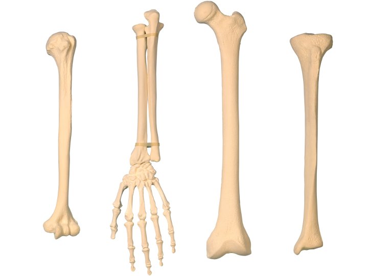
Os
Configurations de test (1)
