Zheyuan Zhou; Fengjie Zhang; Chao Wan
International Journal of Bioprinting 11(2) (2025), 316–335. Published online 15 January 2025. Original abstract: English. Mechanical specimens were cell-free printed hydrogels; recipient mice and cultured chondrocytes are separate biological experiments.
https://doi.org/10.36922/ijb.5230
1 citation · 1 en 2025–2026
Les titres, résumés et légendes des publications sont présentés en anglais.
Current therapies for articular cartilage defects or degeneration, such as osteoarthritis, remain largely unsatisfactory. There are significant clinical demands for the development of more efficient approaches to enhance the repair or regeneration of articular cartilage lesions. In this study, a newly defined alginate- gelatin (A-G) composite bioink was synthesized using the carbodiimide chemistry crosslinking method. Then, the dual-crosslinking (DC) bioscaffolds containing both chemical and ionic networks were fabricated by 3D-bioprinting technique and Ca2+ treatment. Mimicking the mechanical properties of the pericellular matrix of native articular cartilage, the optimized networks formed in the DC bioscaffolds provided a desirable Young’s modulus and geometric constraints, which were superior to that of the single-crosslinking control group. Chondrocytes encapsulated in the 3D printed A-G-DC bioscaffolds exhibited above 95% viability and higher proliferation rate compared to those in the alginate-only group after 7 days of in vitro culture. Chondrogenic differentiation in vitro was improved in the A-G-DC bioscaffolds compared to that of the alginate-only group, indexed by upregulation of chondrogenic marker gene expression, including SOX9, COL2A1, COMP, and ACAN. In subcutaneous transplantation and osteochondral defect models in severe combined immunodeficiency mice, the A-G-DC bioscaffolds exhibited superior chondrogenesis and repair capacity compared to the alginate-only or no-implant control groups. This was evidenced by increased expression of SOX9 and collagen type II (Col II) and higher ICRS II scores. These findings demonstrate that the biomechanically inspired A-G composite bioink and the 3D-printed A-G-DC bioscaffolds possess excellent cell biocompatibility, satisfactory biomechanical properties, and the ability to promote chondrogenesis, making them a promising candidate for enhancing cartilage regeneration in patients with articular cartilage lesions.
Utilisation des équipements
Un Mach-1 v500csst comprimait jusqu’à rupture des échafaudages d’alginate–gélatine imprimés en 3D sans cellules, à 0,0167 mm/s, pour mesurer le module et le point de rupture. L’article décrit aussi des balayages d’amplitude et de fréquence au Mach-1 pour comparer la viscoélasticité des réseaux simples et doubles.
Matériaux (2)
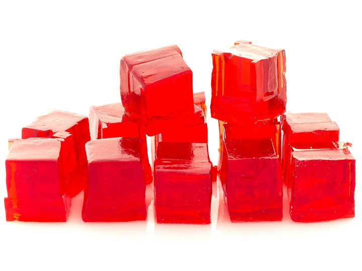
Hydrogel

Matériau par impression 3D
Configurations de test (3)
Compression (confinement non précisé)

Analyse Mécanique Dynamique
