Isabelle Skambath; Jessica Kren; Patrick Kuppler; Steffen Buschschlueter; Matteo Mario Bonsanto
Acta Neurochir 166, 343 (2024), Published: 21 August 2024
https://doi.org/10.1007/s00701-024-06218-4
9 citations · 8 en 2025–2026
Les titres, résumés et légendes des publications sont présentés en anglais.
Abstract
Background
The intraoperative differentiation between tumour tissue, healthy brain tissue, and any sensitive structure of the central nervous system is carried out in modern neurosurgery using various multimodal technologies such as neuronavigation, fluorescent dyes, intraoperative ultrasound or the use of intraoperative MRI, but also the haptic experience of the neurosurgeon. Supporting the surgeon by developing instruments with integrated haptics could provide a further objective dimension in the intraoperative recognition of healthy and diseased tissue.
Methods
In this study, we describe intraoperative mechanical indentation measurements of human brain tissue samples of different tumours taken during neurosurgical operation and measured directly in the operating theatre, in a time frame of maximum five minutes. We present an overview of the Young’s modulus for the different brain tumour entities and potentially differentiation between them.
Results
We examined 238 samples of 75 tumour removals. Neither a clear distinction of tumour tissue against healthy brain tissue, nor differentiation of different tumour entities was possible on solely the Young’s modulus. Correlation between the stiffness grading of the surgeon and our measurements could be found.
Conclusion
The mechanical behaviour of brain tumours given by the measured Young’s modulus corresponds well to the stiffness assessment of the neurosurgeon and can be a great tool for further information on mechanical characteristics of brain tumour tissue. Nevertheless, our findings imply that the information gained through indentation is limited.
Keywords:
Tissue biomechanics, Brain tumour surgery, Indentation measurements, Tissue classification & Elasticity.
Utilisation des équipements
La section de méthode mentionne un appareil Mach-1.
Les détails du protocole sont conservés en anglais dans cette rubrique; consulter la publication originale pour le contexte expérimental complet.
Matériaux (1)
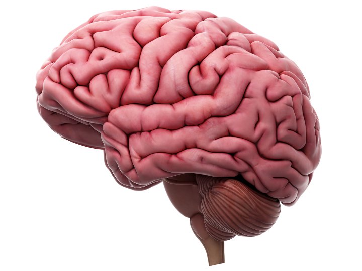
Cerveau
Configurations de test (2)

Cartographie 3D en indentation normale
