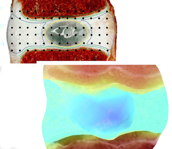
Disk (Nucleus Pulposus)
The nucleus pulposus is the central part of an intervertebral disc.
About this material
The nucleus pulposus is the central part of an intervertebral disc. It is composed of a proteoglycan-water gel embedded in a randomly oriented network of fine collagen fiber and non-collagenous proteins. One of the primary changes in disc degeneration is decreasing proteoglycans and water content within the nucleus pulpus. Knowledge on the mechanical properties of healthy nucleus pulposus will aid in the development of suitable replacement materials and successful disc degeneration treatments.
Test configurations (13)
Unconfined Compression
Indentation (Stiffness)
3D Thickness Mapping
Torsion
Shear

Temperature Control
Mechanical Stimulation (Shear)
Mechanical Stimulation (Compressive)
External Analog Sensors
Camera Feed
Positioning (Manual)
Positioning (X Auto)
Case studies (1)
Related resources (5)
Related literature
Can UVA-light-activated riboflavin-induced collagen crosslinking be transferred from ophthalmology to spine surgery? A feasibility study on bovine intervertebral disc
Ioannis Vasilikos; Graciosa Q. Teixeira; Andreas Seitz; Julia Nothelfer; Julian Haas; Hans-Joachim Wilke; Boris Mizaikoff; Jürgen Beck; Ulrich Hubbe; Cornelia Neidlinger-Wilke
PLoS ONE 16(6): e0252672. https://doi.org/ 10.1371/journal.pone.0252672
5 citations · 2 in 2025–2026
Chitosan-based hydrogels supplemented with gelatine and Link N enhance extracellular matrix deposition by encapsulated cells in a degenerative intervertebral disc environment.
Laboratory for endovascular biomaterials (LBeV), CHUM research centre (CRCHUM), 900 St Denis St, Montreal, QC H2X 3H8, Canada; A Adoungotchodo; LM Epure; F Mwale; S Lerouge
Eur Cell Mater. 2021 May 4;41:471-484. doi: 10.22203/eCM.v041a30. PMID: 33945627.
17 citations · 8 in 2025–2026
Development of an In Vitro Outer Annulus Fibrosus Cartilage Endplate Model and its Response to Dynamic Mechanical Loading
University of Toronto
Intervertebral disc implant
US Patent App. 10195044, 2019
Changes in Free Calcium Alter the Mechanical Properties of the Intervertebral Disc
I. Hadjab; M.P. Grant; L.M. Epure; M. Garon; E. Quenneville; J. Anthoniou; F. Mwale; M.D. Buschmann
International Conference on the Mechanics of Biomaterials and Tissues, Hawaii, USA, 1 2 Dec 2017, Podium O.48. Presented by E. Quenneville
The Bioengineering of Annulus Fibrosus Tissues Using Phenotypically Distinct Inner and Outer Annulus Fibrosus Cells on Oriented Nanofibrous Polyurethane Scaffolds
PhD thesis, University of Toronto, 2017.
0 citations · 0 in 2025–2026
Changes in Collagen and Sulphated Proteoglycan Synthesis by Multilamellated AF Tissues Cultured in vitro on Aligned Nanofibrous Polyurethane Constructs Under Dynamic Compressive Loading
Presentation #331.2 on Fri May 20 from 16:30 to 18:30 in room 511E during the World Congress on Biomechanics, Montreal, May 2016.
Static and Dynamic Compression Application and Removal on the Intervertebral Discs of Growing Rats
Anne‐Laure Ménard; Guy Grimard; Elise Massol; Irène Londono; Florina Moldovan; Isabelle Villemure
Journal of Orthopaedic Research Month 2015, pp.1-9. (2015)
7 citations · 1 in 2025–2026
Load bearing regenerative repairs for the cartilaginous soft tissue of the intervertebral disc
PhD thesis, Queen’s University, August 2014.
0 citations · 0 in 2025–2026
Assessment of mechanical properties of isolated bovine intervertebral discs from multi-parametric magnetic resonance imaging
Maximilien Recuerda; Delphine Périé; Guillaume Gilbert; Gilles Beaudoin
BMC Musculoskeletal Disorders. 2012;13:195.
30 citations · 1 in 2025–2026
Characterization of intervertebral disc mechanical properties and their relationships with quantitative magnetic resonance imaging
Polytechnique Montréal. MASc. 2010
Characterization of the mechanical properties of intervertebral discs and their relationships with quantitative magnetic resonance imaging [English translation]
École Polytechnique de Montréal, Université de Montréal. Master of Applied Science thesis. December 2010.
Tissue engineered nucleus pulposus tissue formed on a porous calcium polyphosphate substrate
Cheryle A. Séguin; Marc D. Grynpas; Robert M. Pilliar; Stephen D. Waldman; Rita A. Kandel
Spine, 29(12), 1299-1306. (2004)
101 citations · 1 in 2025–2026
Towards a Complete Intervertebral Disc: Modulation of the Biochemical and Mechanical Properties of in vitro formed Nucleus Pulposus Tissue
MASc thesis, University of Toronto, 2004.
0 citations · 0 in 2025–2026
A fibril-network-reinforced biphasic model of cartilage in unconfined compression
J. Soulhat; M. D. Buschmann; A. Shirazi-Adl
Journal of Biomechanical Engineering, Jun, 121(3), 340-347. (1999)
250 citations · 9 in 2025–2026
FAQ
How does the "Elastic Model in Indentation" works?+
This tool allows for the computing and visualizing of the shear modulus (MPa) and the Young’s modulus (MPa) from an indentation test performed using a spherical indenter (Figure 1). This model is well adapted for the mechanical characterization of samples (with critical diameter (width) at least 5 times the indenter diameter) bonded on a flat rigid support (at least 10 times stiffer than the sample), for example: any sheet of material lying on a rigid chamber base (limited sliding at the interface can be assumed) or a cartilage layer attached to a thick sub-chondral bone. A user license is required to obtain the results of the elastic model in indentation (Software Add-On, contact Biomomentum to obtain your license).
Results analysis is performed considering the indentation mechanics of an infinitely wide elastic layer bonded to a rigid half-space which has been developed as a model for the layered geometry of cartilage and subchondral bone (Hayes et al., 1972). (Figure 2) shows a schematic representing the modeled situation.
Many parameters need to be specified in order to calculate the different material properties.
1. Indenter Shape is:
- Spherical
- Plane-Ended Cylindrical
2. Indenter radius in mm.
3. Poisson ratio can be selected from a drop-down list (Figure 3)
4. Position @ contact is – defines the position of the left cursor:
- to be curvefit – will automatically find the position contact which will give the smallest mean squared error (mse)
- the value at t0 – will automatically take the position contact at the start of the curve
- the following value (mm) – the user need to specify the value in the corresponding field next to the drop-down list
- cursor position – the user can drag the left cursor to the wanted position contact
5. Sample thickness is the:
- position @ contact – will automatically find the position at contact (position of the left cursor)
- following value (mm) – the user need to specify the value in the corresponding field next to the drop-down list
6. Curvefit is up to – defines the position of the right cursor:
- peak load – will automatically take the position of the peak load
- delta load (unit) – the user need to specify the value of the desired delta load in the corresponding field next to the drop-down list
- delta position (mm) – the user need to specify the value of the desired delta position in the corresponding field next to the drop-down list
The error on the estimation of the best curve representing the stress relaxation curve is calculated using the root of the mean squares (RMS) method and is displayed under “RMSE”. For example, (Figure 4) shows parameters used for the “Normal Indentation” (Mach-1 Motion) function in an indentation testing protocol on ovine articular surfaces. Corresponding results are shown in (Figure 1).
Spherical Indentation Guide – Mach-1 Mechanical Tester – Biomomentum
Mach-1 Analysis – Elastic Model in Indentation – Parameters
Mach-1 Analysis – Elastic Model in Indentation – Poisson ratio choices
Mach-1 Analysis – Elastic Model in Indentation – Cartilage & Subchondral bone
How does the "Poroelastic Model in Confined Compression" feature works?+
This model is well adapted for the mechanical characterization of samples containing a liquid fraction and a solid matrix, for example: cartilage or gel samples. A user license is required in order to obtain the results of the poroelastic model in confined compression (Software Add-On, contact Biomomentum to obtain your license).
Mechanical properties can be obtained from the stress relaxation curves obtained under confined compression such as the aggregate modulus (HA) and the hydraulic permeability (k). The aggregate modulus is a measure of stiffness of the sample at equilibrium while the hydraulic permeability is the ability of the fluid to flow through the cartilage matrix.
For example, (Figure 2) shows parameters used for the “Stress Relaxation” function in a confined compression testing protocol on human cartilage disks. Corresponding results are shown in (Figure 1).
Mach-1 Analysis – Poroelastic Model in Confined Compression – Parameters
Mach-1 Analysis – Poroelastic Model in Confined Compression – Example
How does the "Poroelastic Model in Unconfined Compression" feature works?+
The nonhomogeneous composite model (i.e. linear fibril-network-reinforced biphasic model) is an extension of the previously developed isotropic homogeneous biphasic model, and was achieved by reinforcing the latter with a network of nonlinear fibrils distributed homogeneously in the r, θ, z directions and which resist extension only (Figure 2). The isotropic matrix is described by the drained elastic constants Em (Young’s modulus), vm (Poisson’s ratio) and a hydraulic permeability k. The fibril network is described by the Young’s modulus of individual fibrils ef which, combined with a fibril volume fraction χ (fibrils volume/ total-volume), results in an equivalent tensile modulus of the fibril network, Ef = 1/3 χ ef (since 1/3 of the fibrils are oriented in each of three mutually orthogonal directions).
For example, (Figure 3) shows parameters used for the “Stress Relaxation” function in an unconfined compression testing protocol on ovine cartilage disks. Corresponding results are shown in (Figure 1).
Mach-1 Analysis – Poroelastic Model in Unconfined Compression – Parameters
Mach-1 Analysis – Poroelastic Model in Unconfined Compression – Cartilage
Mach-1 Analysis – Poroelastic Model in Unconfined Compression – Example
How does the "Sinusoid" model works?+
This feature is used to obtain the Frequency, Amplitude and Offset of a sinusoidal signal. A blue line displays the theoretical sinusoid with the Frequency, Amplitude and Offset obtained (Figure 1). The values of the Frequency, Amplitude and Offset are indicated in the "Results" field. The units vary according to the graph’s axes.
This feature can also be used for sinusoidal stimulus-response analysis. Sinusoidal stimulus shall be selected on the x-axis and the sinusoidal response on the y-axis. The Modulus is the ratio of the response Amplitude over the stimulus Amplitude. The Phase is the phase difference between the response and the stimulus. The Modulus’ is computed as the Modulus*Cos(Phase) and the Modulus” is computed as the Modulus*Sin(Phase). A blue line displays the theoretical sinusoidal stimulus-response with the Frequency, Modulus and Phase obtained (Figure 2). The values of the Modulus and Phase are indicated in the “Results” field. The units vary according to the graph’s axes.
One use for this feature would be to compute material properties by multiplying the Modulus obtained by the appropriate geometric factors. For example, the Young’s Modulus can be obtained during uniaxial compression or tension on cylindrical sample with t/pi*r2 as the geometric factor, t is the thickness and r is the radius of the sample.
Mach-1 Analysis – Sinusoid – Example for stimulus-response analysis
What is an aggregate modulus?+
How does the creep function works?+
What is viscoelasticity?+
LAB MODULE – Torsion of Disk-Shaped Samples – Student Version
What is the elasticity?+
LAB MODULE – Unconfined Compression of Hydrogel Disks – Student Version