Changes in Free Calcium Alter the Mechanical Properties of the Intervertebral Disc
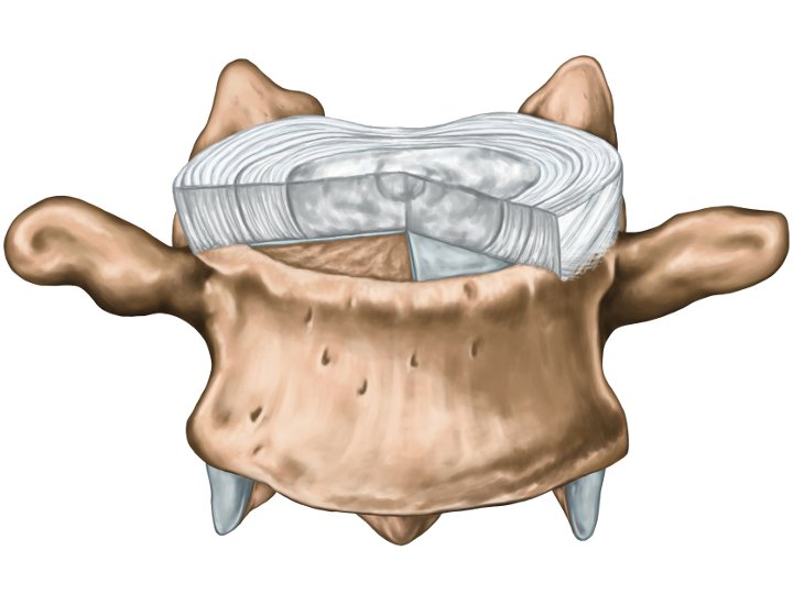
Since we recently discovered that increased free calcium (Ca2+) concentration in the human intervertebral disc (IVD) leads to matrix degradation (Grant et al. ECM 2016), we hypothesized that elevations in Ca2+ also impair IVD mechanics. The aim of this study was to determine the effect of free Ca2+ on the mechanical properties of the IVD.
IVDs were isolated from 25-month-old bovine tails and incubated for 4 weeks with either 1mM or 5mM Ca2+. IVDs without calcium supplementation were considered as control. Using the Mach-1 mechanical tester (Biomomentum Inc, Canada), each IVD was subjected to compressive dynamic mechanical testing (DMA) to assess its bulk Young's modulus (E*) for frequencies ranging from 0.01Hz to 5Hz using 5% strain amplitude. Subsequently, to assess the compartmental stiffness, the IVDs were divided in two halves along the spinal cord, and the central section of each half was assessed using an automated spherical indentation technique (R=3.2mm, 15 measures/cm2, amplitude=0.5mm). Stiffness maps were generated.
After a 4-week incubation period in 1mM or 5mM Ca2+, E* decreased significantly compared to the control. Stiffness of IVD's compartments remained almost unchanged when compared to the control for low Ca2+ concentration (1mM), while it reached significance (p<0.0001) at the high
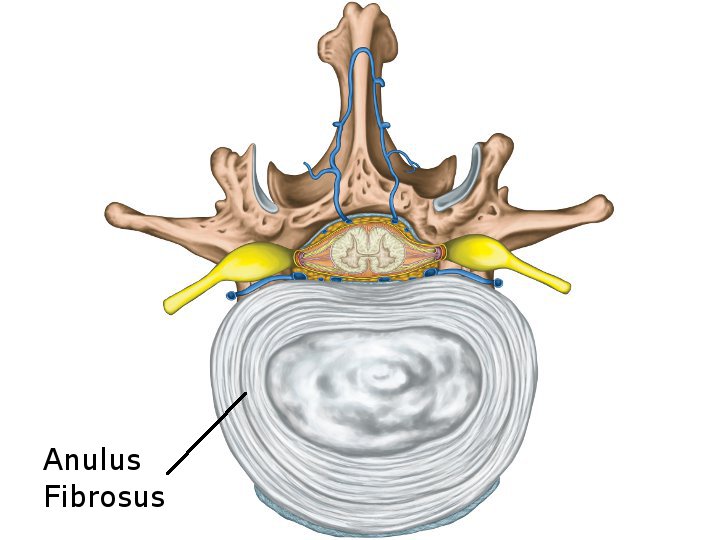
concentration (5mM). The stiffness decreased in annulus fibrosus (AF) from 0.30±0.02MPa to 0.06±0.01MPa. However, for both nucleus pulposus (NP) and endplates (EP), tissues stiffness increased from 0.030±0.001MPa to 0.060±0.002MPa and from 1.45±0.15MPa to 2.5±0.07MPa,
respectively.
concentration (5mM). The stiffness decreased in annulus fibrosus (AF) from 0.30±0.02MPa to 0.06±0.01MPa. However, for both nucleus pulposus (NP) and endplates (EP), tissues stiffness increased from 0.030±0.001MPa to 0.060±0.002MPa and from 1.45±0.15MPa to 2.5±0.07MPa,
respectively.
High Ca2+ concentration alters IVD's bulk and compartmental mechanical properties. These results are consistent with our previous findings suggesting that increasing the calcium levels resulted in increases in the mineralisation of EP, and decrease the diffusion of nutrient into the disc. Consequently, assessing mechanical properties of each IVD's compartment may provide a better understanding of its bulk mechanical behavior and its integrity during degeneration.
Adapted from: I. Hadjab, M.P. Grant, L.M. Epure, M. Garon, E. Quenneville, J. Anthoniou, F. Mwale and M.D. Buschmann. Changes in Free Calcium Alter the Mechanical Properties of the Intervertebral Disc. International Conference on the Mechanics of Biomaterials and Tissues, Hawaii, USA, 12Dec2017, Podium O.48.
Figures

Figure 1 — Show caption
Measurement grid and color map illustrating regional mechanical properties of an intervertebral disc.

Figure 2 — Show caption
Disc stiffness maps after four weeks under control and calcium-supplemented conditions.

Figure 3 — Show caption
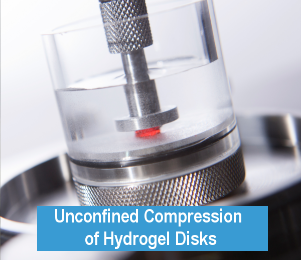
Compression test setup and dynamic Young’s modulus of discs under different calcium conditions.

Figure 4 — Show caption
Stiffness of the endplates, annulus fibrosus and nucleus pulposus under the conditions shown.
Products (3)


Accessories (20)



Test configurations (5)

Test configuration
3D Normal Indentation Mapping

Test configuration
Unconfined Compression

Test configuration
Indentation (Stiffness)

Test configuration
Camera Feed

Test configuration