Tim Finkenstaedt; Reni Biswas; Nirusha A. Abeydeera; Palanan Siriwanarangsun; Robert Healey; Sheronda Statum; Won C. Bae; Christine B. Chung
Invest Radiol. 2019 Jun. 54(6):349-355.
https://doi.org/10.1097/RLI.0000000000000547
21 citations · 4 en 2025–2026
Les titres, résumés et légendes des publications sont présentés en anglais.
OBJECTIVES:
In human menisci, we aimed to investigate whether calcium pyrophosphate crystal deposition (CPPD) affects biomechanical and quantitative MR properties, and their zonal distribution.
MATERIALS AND METHODS:
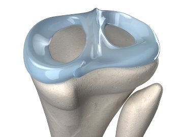
From 9 cadaveric knees, sectioned triangular meniscus pieces were harvested. Samples were classified into « normal » or « CPPD » groups based upon visual inspection. Micro computed tomography scan verified CPPD. Using magnetic resonance imaging, ultrashort echo time (UTE) T2* and spin echo (SE) T2, quantitative values in 3 zones (red, red-white, and white) were determined. Using biomechanical test, indentation forces in the same zones were determined. Effects of CPPD and meniscal zone on indentation force and quantitative MR values were compared.
RESULTS:
On UTE MRI scans, CPPD-affected menisci exhibited punctate dark regions, found mostly (92%) in avascular white and red-white zones. Indentation forces were significantly higher for CPPD samples in the red-white (all P < 0.02) and white (all P < 0.004) zones but not in the vascular red zone (all P > 0.2). Similarly, UTE T2* red zone values were similar between both groups (~6.6 milliseconds, P = 0.8), whereas in the red-white and white zones, CPPD samples had significantly lower values (~5.1 milliseconds, P = 0.005 to 0.007). In contrast, SE T2 values showed no difference with CPPD (P = 0.12 to 0.16). UTE T2*, but not SE T2, correlated significantly with indentation force (R = -0.29, P = 0.009).
CONCLUSIONS:
Dark CPP deposits were detectable on UTE images featuring high signal intensity from surrounding meniscal tissue. Preliminary results indicate that CPP deposits were almost exclusively found in the avascular zones. Compared with normal, CPPD menisci featured higher indentation stiffness and lower UTE T2* values in the affected zones.
Utilisation des équipements
Des échantillons de ménisque humain cadavérique, avec ou sans dépôts de cristaux de pyrophosphate de calcium, ont été soumis à des essais d’indentation par zone, parallèlement à une IRM quantitative. Les forces d’indentation ont été mesurées dans les zones rouge, rouge-blanche et blanche. Le résumé disponible n’identifie pas l’appareil mécanique et ne précise pas suffisamment le protocole pour vérifier le rôle d’un Mach-1.
Configurations de test (1)