Heta Orava; Petri Paakkari; Jiri Jäntti; Miitu K. M. Honkanen; Juuso T. J. Honkanen; Tuomas Virén; Anisha Joenathan; Petri Tanska; Rami K. Korhonen; Mark W. Grinstaff; Juha Töyräs; Janne T. A. Mäkelä
Wiley Online Library, Journal of ORS, published: 18 August 2023
https://doi.org/10.1002/jor.25683
7 citations · 5 en 2025–2026
Les titres, résumés et légendes des publications sont présentés en anglais.
Abstract
Cartilage and synovial fluid are challenging to observe separately in native computed tomography (CT). We report the use of triple contrast agent (bismuth nanoparticles [BiNPs], CA4+, and gadoteridol) to image and segment cartilage in cadaveric knee joints with a clinical CT scanner. We hypothesize that BiNPs will remain in synovial fluid while the CA4+ and gadoteridol will diffuse into cartilage, allowing (1) segmentation of cartilage, and (2) evaluation of cartilage biomechanical properties based on contrast agent concentrations. To investigate these hypotheses, triple contrast agent was injected into both knee joints of a cadaver (N = 1), imaged with a clinical CT at multiple timepoints during the contrast agent diffusion. Knee joints were extracted, imaged with micro-CT (µCT), and biomechanical properties of the cartilage surface were determined by stress-relaxation mapping. Cartilage was segmented and contrast agent concentrations (CA4+ and gadoteridol) were compared with the biomechanical properties at multiple locations (n = 185). Spearman's correlation between cartilage thickness from clinical CT and reference µCT images verifies successful and reliable segmentation. CA4+ concentration is significantly higher in femoral than in tibial cartilage at 60 min and further timepoints, which corresponds to the higher Young's modulus observed in femoral cartilage. In this pilot study, we show that (1) large BiNPs do not diffuse into cartilage, facilitating straightforward segmentation of human knee joint cartilage in a clinical setting, and (2) CA4+ concentration in cartilage reflects the biomechanical differences between femoral and tibial cartilage. Thus, the triple contrast agent CT shows potential in cartilage morphology and condition estimation in clinical CT.
Methods
µCT imaging and biomechanical mapping
The samples were thawed, and any remaining contrast agent was washed out of the cartilage over a period of 4 days as described in the Supporting Information: Articular cartilage thickness was obtained from µCT images by using a custom-made MATLAB code. The imaging was conducted in the air with Nikon XT H 225 µCT scanner (Nikon Metrology Europe), with an isotropic voxel size of 50 µm × 50 µm × 50 µm, tube voltage of 120 kVp, tube current of 167 µA, tube power of 20.0 W, and filtering with 0.5 mm aluminum filter. Mapping of the biomechanical properties was conducted using a multiaxial mechanical tester Mach-1 v500css (MA006; Biomomentum Inc.) with a multiaxial 17 N load cell (MA232; Biomomentum Inc.). Details on the stress–relaxation protocol can be found in Supporting Information: Section S5. For each measured location, instantaneous and equilibrium Young's moduli were calculated as described in Supporting Information.
Utilisation des équipements
Le Mach-1 mesurait les propriétés mécaniques du cartilage par un protocole de cartographie avec relaxation des contraintes.
Matériaux (2)

Matériau
Cartilage articulaire
Matériau
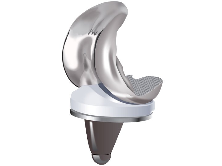
Prothèse (genou)
Configurations de test (2)

Configuration de test
Indentation (Raideur)
Configuration de test