Brenda Ogle; Sophie Givens; Abygail Andebrhan; Ruchen Wang; Xiangzhen Kong; Taylor Rothermel; Sanaz Hosseini; An Xi; Mohammed Shameen; Andrea Torniainen; Samuel Boland; Maya Johnson; Natalia Mancipe; Bhairab Singh; Samuel Dudley; Patrick Alford; Elena Tolkacheva; Jop van Berlo
PREPRINT (Version 1) available at Research Square , Published: 11 June 2024
https://doi.org/10.21203/rs.3.rs-4450763/v1
0 citations · 0 en 2025–2026
Les titres, résumés et légendes des publications sont présentés en anglais.
Abstract
Accumulating evidence in vitro indicates that the exposure of human induced pluripotent stem cell-derived cardiomyocytes (hiPSC-CM) to adult post-natal environmental conditions is not sufficient to attain maturation. Here we instead mimic the progressive embryonic ventricular environment via co-culture of hiPSC-CM with epicardial cells and epicardial-derived cells in two-dimensional (2D) direct co-culture and 3D engineered heart tissues (EHT). When epicardial cells were combined with CM in 2D direct co-cultures the percentage of proliferating CM increased at the same time as stark electrophysiologic improvements in calcium handling.
Notably, no increase in single-cell CM force generation was observed, as determined using traction force microscopy. Single-cell transcriptomics revealed a significant shift in the bulk CM population in epicardial-CM co-cultures as characterized by more fetal-like myofilament isoforms but with enhanced pathways associated with cardiac conduction and ventricular development. While co-culture of epicardial cells in the 3D-EHT model showed limited proliferation but a similar improvement in CM electrophysiologic function. These results indicate that certain aspects of CM maturation are enhanced, especially cardiac conduction, by epicardial cells while an embryonic phenotype is maintained (i.e., proliferation and myofilament
isoforms). Next, consistent with developmental progression and the initiation of epicardial-to-mesenchymal transition (EMT), epicardial-derived fibroblasts were added to the EHTs containing epicardial cells. Interestingly, we observed significant myofilament maturation and increased force generation by more than an order of magnitude. Taken together, our results support the importance of mimicking developmental progression to slowly taper proliferation while gradually driving the maturation of hiPSC-CM to achieve robust in vitro function at the cell and tissue scale.
Utilisation des équipements
Le Mach-1 enregistrait les forces de contraction de tissus cardiaques reconstruits stimulés électriquement à des déformations en traction imposées.
Matériaux (1)
Matériau
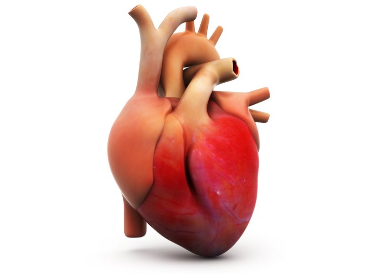
Coeur (génie tissulaire)
Configurations de test (2)

Configuration de test
Tension

Configuration de test