Ying Betty Li; Caroline Sodja; Marina Rukhlova; Jordan Nhan; Joshua J.A. Poole; Harry Allen; Selam Yimer; Ewa Baumann; Erin Bedford; Hannah Prazak; Will J. Costain; Sangeeta Murugkar; Jean-Philippe St-Pierre; Leila Mostaço-Guidolin; Anna Jezierski
SSRN preprint, 2022.
https://doi.org/10.2139/ssrn.4068146
3 citations · 1 en 2025–2026
Les titres, résumés et légendes des publications sont présentés en anglais.
Despite recent advances in 3D bioprinting technologies, the biofabrication of complex vascular network architectures has remained a key challenge in engineering tissues. In this study, we leveraged a novel microfluidic based 3D bioprinting technology and alginate-collagen type I (AGC)-based bioinks, to develop a 3D bioprinting strategy to enable the biofabrication of complex vascular networks comprised of SV40 transformed adult rat brain endothelial cell (SV-ARBEC)-laden hydrogel rings with mechanical properties relevant for neurovascular tissue engineering applications. The bioprinted SV-ARBECs retained high cellular viability, at high tissue level cell densities, and formed spontaneous endothelial vascular networks reminiscent of anisotropic tissue-like structures. The vascular network formation was accompanied by extracellular matrix remodeling (ECM) remodelling, evaluated by second harmonic generation (SHG) microscopy, confirming sequential SV-ARBEC mediated collagen type I fiber deposition and reorganization. This novel 3D microfluidic bioprinting technology and biocompatible AGC hydrogel fiber rings enabled robust SV-ARBEC angiogenesis and vascular network formation which are key towards advancing applications in vascular research and development, disease modeling, drug discovery and tissue engineering applications.
Utilisation des équipements
Un Mach-1 de Biomomentum a caractérisé des disques moulés d’hydrogel à base d’alginate avec une cellule de charge uniaxiale de 1,5 N et un indenteur sphérique de 1 mm. Les échantillons immergés ont subi une indentation à 25 % de déformation, puis 60 secondes de relaxation. Les données ont été analysées selon le modèle de Hayes.
Matériaux (1)
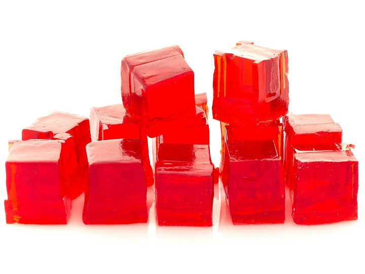
Hydrogel
Configurations de test (2)
