Kolaczek S; Changoor A; Hurtig M; Gordon K; Getgood A
Annual Meeting of the ORS, New Orleans, Louisiana, USA, March 15-18, 2014.
Les titres, résumés et légendes des publications sont présentés en anglais.
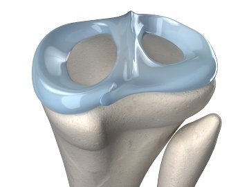
A major risk factor for knee osteoarthritis (OA) is meniscal injury or excision. Surgeons now advocate for meniscal repair or allografts rather than excision with this knowledge in hand. Clinical assessment of the meniscal load sharing is limited to meniscus position by MRI, so the efficacy of replacement and repair must be done in cadaveric knees. In the past, pressure sensitive film and mechanical transducers have been used [3, 4] but our laboratory has measured regional strain in menisci using CT imaging and small implanted beads. One problem is that determining the volume of the meniscus requires segmentation in MRI or CT which is not accurate, particularly near capsular attachments. We hypothesized that contrast enhancement using heavy metals may allow automated segmentation of the volume on a CT image, allowing assessment of meniscal repair strategies from CT scans. The objectives of this study were to investigate whether indentation properties of the meniscus change after exposure to mercury, and to determine whether mercury exposure improves visualization of the entire meniscus in micro-computed tomography (micro-CT).
Utilisation des équipements
Le Mach-1 réalisait des indentations de ménisques porcins intacts. La normale locale était calculée à partir de mesures de détection de surface environnantes.
Article et documents
Document
Programme de conférence (PDF)
Matériaux (1)
Matériau
Ménisque
Configurations de test (3)

Configuration de test
Cartographie 3D en indentation normale

Configuration de test
Cartographie 3D d'épaisseur

Configuration de test