Boeun Hwang
Georgia Institute of Technology and Emory University, PhD dissertation, May 2025 (cover). Repository metadata date 19 February 2025 is not used as publication day. Original complete English summary: PDF pp.13–14 and repository description. Methods 3.1.2 and 3.1.7: PDF pp.50 and 53. Human detail refers to HUVECs in the patch, not native human heart. Probe dimension copied without assuming radius or diameter. Thesis and associated journal article remain distinct bibliographic documents.
Les titres, résumés et légendes des publications sont présentés en anglais.
With recent advances in the field of tissue engineering, cardiac patch devices have shown great promise to restore the heart function and structure following myocardial infarction (MI). Cardiac patches can provide mechanical support to the MI tissue, while enabling sustained and targeted delivery of regenerative factors to the infarct site to promote healing. Despite the significant progress made towards fabrication of functional cardiac patches, many of the current patch devices face challenges, such as the lack of vasculature and insufficient biomolecular cues to boost regenerative effects. This thesis aims to develop a new generation of multifunctional cardiac patches with perfusable vasculature. Leveraging 3D bioprinting technology, patch constructs with a bifurcated vasculature were fabricated and endothelialized using human umbilical vein endothelial cells (HUVECs). The endothelialized vascular patches supported 3D culture of HUVECs and maintained a healthy endothelium after perfusion culture. Additionally, we aimed to overcome the limited regenerative capacity of cardiomyocytes (CMs) by incorporating pro-proliferative molecules, i.e., follistatin-like 1 (FSTL1) and CHIR99021, in the constructs. The effect of FSTL1 and CHIR99021 on human induced pluripotent stem cell (hiPSC)-derived CM proliferation was assessed in 2D and 3D cultures. Finally, the synergistic effect of FSTL1 and CHIR99021 was studied within our 3D bioprinted vascular patch and in a rat model of acute MI. The delivery of CHIR+FSTL1-loaded cardiac patch to the infarcted heart significantly improved cardiac function and reduced scarring. Furthermore, enhanced angiogenesis and CM proliferation were observed after the CHIR+FSTL1 patch treatment. Through integration of 3D bioprinting and regenerative medicine, the cardiac patches engineered in this thesis offer an outstanding therapeutic potential to treat myocardial injuries.
Utilisation des équipements
Un Mach-1 mesurait le module de patchs cardiaques GelMA–gélatine–fibrine contenant des cellules endothéliales humaines, par microindentation avec une sonde sphérique de 300 µm. Le calcul utilise les courbes de décharge. La fonction normale 3D n’est pas documentée; l’implantation chez le rat est une expérience distincte.
Article et documents
Consulter la source
Matériaux (2)
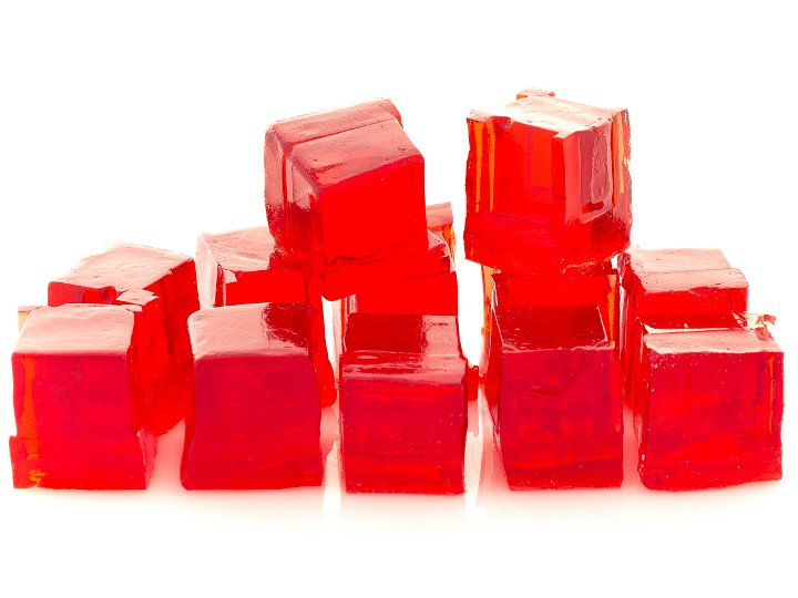
Hydrogel
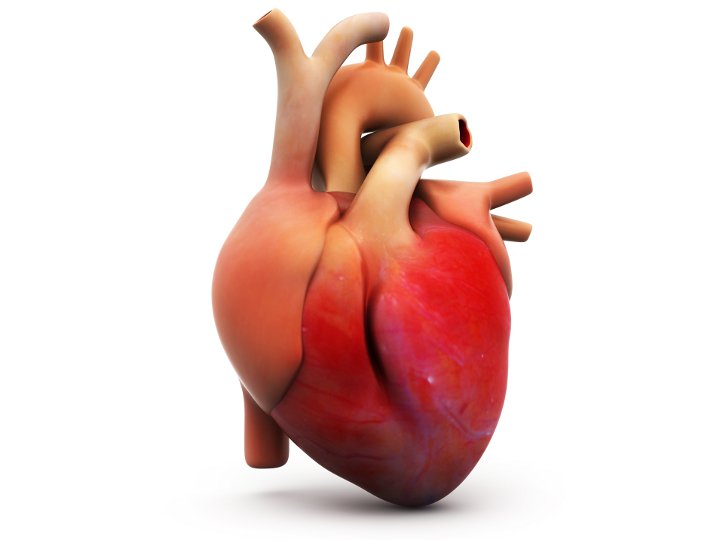
Coeur (génie tissulaire)
Configurations de test (1)
